Case of the week 3.12.18
Publication Date: 2018-03-09
History
2 year old male castrated rottweiler, vomting. Was treated with NSAIDs
7 images
Findings
Orthogonal radiographs of the abdomen are available for interpretation
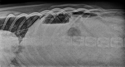
The serosal margin detail is diffusely mildly decreased, with fluid streaking of the peritoneal fat. Numerous small, variably sized gas foci are noted within the peritoneal cavity, not contained within the gastrointestinal tract.
The presence of free peritoneal gas is confirmed on the horizontal beam projection where a moderate amount of free peritoneal gas is seen caudal to the pylorus.
The liver, spleen, kidneys and urinary bladder are normal. The stomach contains a small amount of gas and is minimally distended.
Gas is seen within the pylorus on the left lateral and VD views. A few small, variably shaped and sized mineral opacities are noted within the stomach, the largest of which measures approximately 1.5 cm and is triangular in shape. These mineral opacities vary in position between various projections. Mixed soft tissue and gas opaque granular material is noted within several small intestinal segments. A few pinpoint, mineral opacities are also noted within a few small intestinal segments.
There is marked segmental gas dilation of an intestinal segment in the right cranial abdomen which may represent a portion of the colon or may represent a segment of small intestine. The descending colon contains a small amount of mixed soft tissue and gas opaque granular fecal material.
The included thoracic structures are normal. There is mild bilateral coxofemoral periarticular new bone formation.
Diagnosis
- Pneumoperitoneum and peritonitis, most likely secondary to gastrointestinal perforation. Possible etiologies include perforation of a gastric ulcer (considering the reported history of NSAID administration) or a small intestinal foreign body. If the gas distended intestinal segment in the right cranial abdomen represents a small intestinal segment, this segment is excessively distended and would support small intestinal ileus. Material within several small intestinal segments may represent foreign material although the presence of ingesta cannot be ruled out. Exploratory laparotomy is recommended.
Notes
Due to financial constraints, the owner elected to have the exploratory laparotomy performed at their referring veterinarian. The patient was lost to follow up.
Files
