Case of the week 2.26.18
Publication Date: 2018-02-26
History
1 year old cat. 1 week history of weight loss and progressive anemia.
3 images
Findings
Orthogonal radiographs of the abdomen.
There is mild decreased serosal detail of the retroperitoneal space characterized by mild curvilinear soft tissue streaking of the retroperitoneal fat.
The kidneys are bilaterally moderately enlarged and maintain normal margination.
The liver extends mildly beyond the costal arch but remains sharply marginated. The gastrointestinal tract, spleen and urinary bladder are normal. The included musculoskeletal structures are normal.
Bilateral renomegaly with suspect retroperitoneal effusion is most suggestive of an infiltrative neoplasia such as lymphoma. Given the young age of the patient, acute pyelonephritis or feline infectious peritonitis may also be considered but is considered less likely.
Mild hepatomegaly may be patient variant, however, diffuse infiltration (such as from lymphoma) cannot be ruled out.
Diagnosis
Abdominal ultrasound was performed.


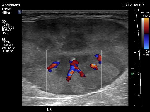
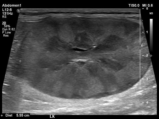
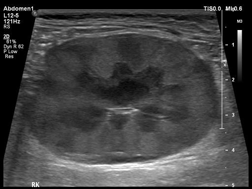
Bilaterally the kidneys are round hyperechoic heterogeneous and there is an hypoechoic subcapsular rim. The retroperitoneal space is diffusely enlarged and hyperechoic. There is mild abdominal lymphadenopathy adjacent to the ileocolic junction. The slpeen was also mottled.
Discussion
The cat was diagnosed with FIP on renal biopsy and subsequently on necropsy.
Files