Case of the week 12.4.17
Publication Date: 2017-12-04
History
13 year old American domestic shorthair. Chronic hypokalemia, polyuria, polydyspia.
3 images
Findings
Orhtogonal radiographs of the abdomen are available for interpretation.
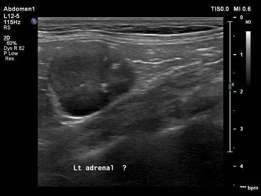
The serosal margin detail is normal. An approximately 2.7 cm round mass is seen within the cranial aspect of the retroperitoneal space, cranial to the left kidney.
The liver, spleen, kidneys, and gastrointestinal tract are within normal limits.
The cardiac silhouette is mildly enlarged on the lateral views. The aorta has an undulating appearance. The pulmonary vasculature and caudal vena cava are normal. There are no abnormalities associated with the visible aspect of the pulmonary parenchyma. A thin pleural fissure line is seen within the caudoventral aspect of the thorax on the right lateral view and minimally on the left lateral view.
Mild rounding of the lumbodiaphragmatic recess of the lung lobes is seen. Mild osseous remodeling is associated with the coxofemoral joints bilaterally.
Diagnosis
- Left adrenal gland mass; given the reported hypokalemia a functional adenoma/adenocarcinoma resulting in hyperaldosteronism is suspected.

- Cardiomegaly suggestive of cardiomyopathy. No evidence of congestive heart failure. Redundant aorta may be seconary to systemic or age related changes. Thin pleural fissure lines are suggestive of a small amount of pleural effusion. Three view thoracic radiographs are recommended for further evaluation and for metastatic check.
Files