Case of the week 11.13.17
Publication Date: 2017-11-13
History
1 year old male castrated golden retriever. Intermittent vomiting and diarrhea for the last 10 months.
5 images
Findings
Orthogonal radiographs of the abdomen are available for interpretation.
There is moderate decrease serosal detail of the peritoneal cavity.
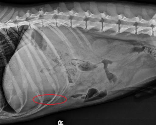
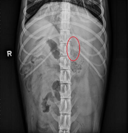
Superimposed with the ventral aspect of the liver and the costal cartilages on the lateral views, there is a well-defined linear gas lucency approximately 4.5cm in length and 0.3 cm in width. This linear lucency is correlated on the VD view, to a similar gas lucency just to the left of T13.
The stomach contains a mixture of gas and heterogenous soft tissue material. On the left lateral the duodenum is gas-filled and there are small outpoucing of its lumen, consistent with Peyer’s patches. The small intestines have a bunched appearance within the mid ventral abdomen. The majority of the small intestines are soft tissue opaque however there are a couple segments which contain gas and are irregularly-shaped, most notable on the right cranial lateral view.
The spleen is moderately diffusely enlarged. The kidneys are normal. The margins of the urinary bladder and caudal margins of the liver cannot clearly be identified due to the decrease serosal detail. The included osseous structures are normal.
Diagnosis
The described linear lucency does not appear to be contained within the gastrointestinal tract and may represent either a foreign body or gas tract.
The decrease serosal detail may be due to peritonitis and/or peritoneal effusion.
- Splenomegaly is most likely secondary to sedation.
Discussion
Abdominal computed tomography and ultrasound were performed.
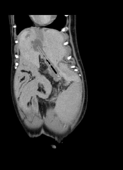
There were 3 pieces of wood within the abdomen, the largest one embedded in the hepatic parenchyma is seen on the dorsal reconstruction of the CT below.
The dog also had multiple associated hepatic abscesses. The patient underwent surgery, the foreign bodies were removed and the abscesses drained.


Files