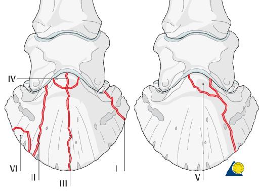
P3 fracture
Publication Date: 2016-12-21
Details
Service Radiology
Modality: Radiographs
Species: Equine
Area: Limb
History
11 year old american quarter horse. Acutely lame a month ago, very sensitive to hoof testers across the heel.
7 images
Findings
Left front foot: Multiple lateral, DP, high oblique DP, and navicular skyline views are available.
There is a type II non-displaced fracture of the distal phalanx along the medial aspect. The fracture gap is moderately widened measuring up to approximately 4.5mm at the solar margin of P3. The fracture extends into the medial aspect of the distal interphalangeal joint. The fracture margins are smoothly rounded suggesting chronicity.
here is a slightly increased number of synovial invaginations in the navicular bone. The remaining skeletal structures are normal.
Diagnosis
Chronic type II fracture of the distal left front phalanx as described with articular involvement of the distal interphalangeal joint.
Mild navicular degeneration may be an incidental finding given the concurrent distal phalangeal fracture.
There are at least 6 types of classified P3 fractures depending on their location and their potential joint involvement.

Notes
Files