Panosteitis
Publication Date: 2016-12-21
Details
Service Radiology
Modality: Radiographs
Species: Canine
Area: Limb
3 images
Findings
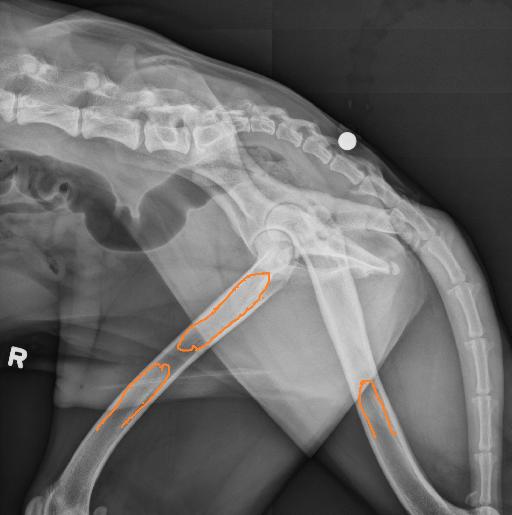
A right lateral view, a right lateral view with the legs spread apart and a VD view are available.
There are multifocal patchy areas of increased opacity throughout the medullary cavity of both femora.
The coxofemoral joints are within normal limits. There is a transitional lumbosacral vertebra with sacralization of the left and lumbarization of the right side.
Diagnosis
Findings are consistent with panosteitis. No evidence of hip dysplasia based on these radiographs. Transitional lumbosacral vertebra may be incidental at this point but may on occasion be associated with an increased risk for coxofemoral degenerative joint disease and cauda equina compression syndrome.

Notes
Files