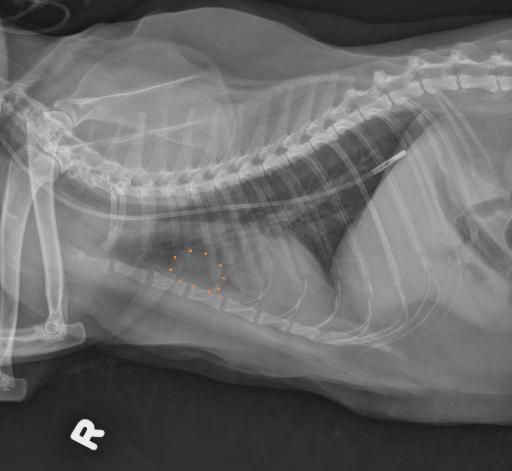
13 year old mixed breed cat. Female spayed. Tube placement, history of triaditis and anorexia
Publication Date: 2016-12-16
History
13 year old mixed breed cat. Female spayed. Tube placement, history of triaditis and anorexia
3 images
Findings
There is widening of the cranial mediastinum immediately cranial to the heart. This region appears soft tissue on the lateral views.
The cardiac silhouette, pulmonary vasculature, and pulmonary parenchyma are normal. There is a nasogastric tube which extends along the plane of the esophagus. The distal end lies superimposed with the diaphgramatic margin. The skeletal structures are normal.
Diagnosis
Focal opacity in the cranial mediastinum likely represents an incidental branchial cyst. Otherwise normal thorax. Feeding tube.

Pathophysiology
Branchial cysts are incidental cyst arising from the remnants of branchial pouch epithelium. They are usually encountered in old animals; however, they have been identified in young animals.
Files