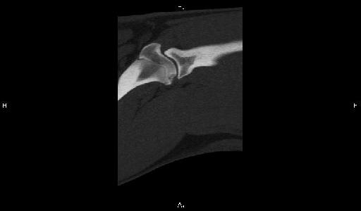
OC shoulder
Publication Date: 2016-12-09
Details
Service Radiology
Modality: Radiographs
Species: Canine
Area: Limb
History
1 year old female spayed Bernese Mountain Dog. History of forelimb lameness more marked on the left side. Bilateral hip pain on the orthopedic exam
6 images
Findings
Orthogonal radiographs of the pelvis and both shoulders are available for interpretation.
Overall the pelvis is considered to be within normal limits.
Bilaterally but most marked on the left side there is flattening of the caudal aspect of the humeral head. Furthermore on the left side this flattening is also irregular. There is no evidence of osseous fragment within the joint space.
Quizz
- What are the most common sites of osteochondrosis in the dog ?Caudal aspect of the proximal humeral head
Yes :)Distomedial aspect of the humeral trochlea
Yes :)Lateral and medial femoral condyles
Yes :)Medial and lateral trochlear ridges of the talus.
Yes :)All of the above
Yeah !
Diagnosis
Bilateral shoulder osteochondrosis most marked on the left side without evidence of joint mice.

Sagital reconstruction of the shoulder joint illustrates the osteochondritic changes in the caudal aspect of the humeral head.
Radiographically normal pelvis. No underlying cause for the pain was found.
Notes
Files