NUPA
Publication Date: 2016-11-18
Details
Service Radiology
Modality: Radiographs
Species: Canine
Area: Limb
4 images
Findings
Orthogonal radiographs of the right elbow as well as flexed lateral view are available for interpreation.
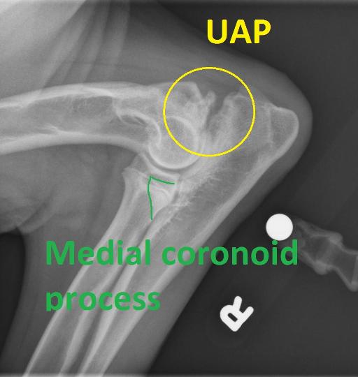
At the level of the anconeal process, there is an irregularly marginated triangularly shaped osseous fragment. The fragment is separated from the remainder of the ulna. The anconeal process and the adjacent ulna are sclerotic. Surrounding the elbow joint there is moderate intracapsular soft tissue swelling. Subjectively the medial coronoid process is blunted and mildly sclerotic. There is mild to moderate osseous remodeling of the medial aspect of the distal humerus.
Diagnosis
Changes are consistent with elbow dysplasia with ununited anconeal process and fragmented coronoid process. Secondary degenerative changes graded as mild to moderate and intracapsular effusion.
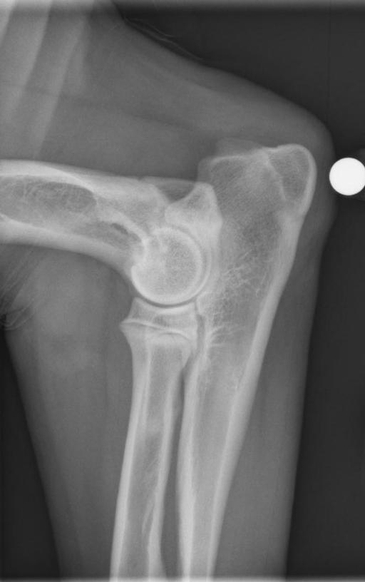
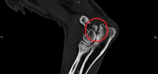
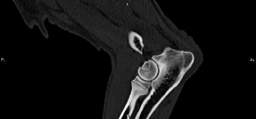
The contralateral limb was normal. The patient underwent CT and the radiological findings were confirmed.


Flexed lateral of the affected elbow and contralatearl elbow for comparison.


CT sagital images to illustrate the non united anconeal process. Contralateral elbow for comparison
Notes
Files