Extraskeletal osteosarcoma and hypertrophic osteophathy
Publication Date: 2016-08-15
3 images
Findings
Orthogonal radiographs of the thorax are available for interpretation.
In the caudal aspect of the left hemithorax there is an ill-defined soft-tissue mass resulting in deviation of the left mainstem bronchus towards the right side. Most appreciated on the lateral views there is amorphous mineralization superimposed with that area. The mass silhouettes with the caudal aspect of the heart. On the lateral views, caudo-dorsally to the mainstem bifurcation there is a well-defined rounded soft tissue mass which extends up to the ventral aspect of the vertebral body. On the right side, the 5th-7th ribs are chronically fractured and misaligned with presence of callus. An additional 1 cm mineral opaque nodule is visible within the fourth/fifth intercostal space on both lateral views. There is a discrete smooth soft tissue nodule located in the right caudal lung lobe that measures approximately 2.3cm in diameter, seen on the left lateral view and VD view under the 9th right rib.
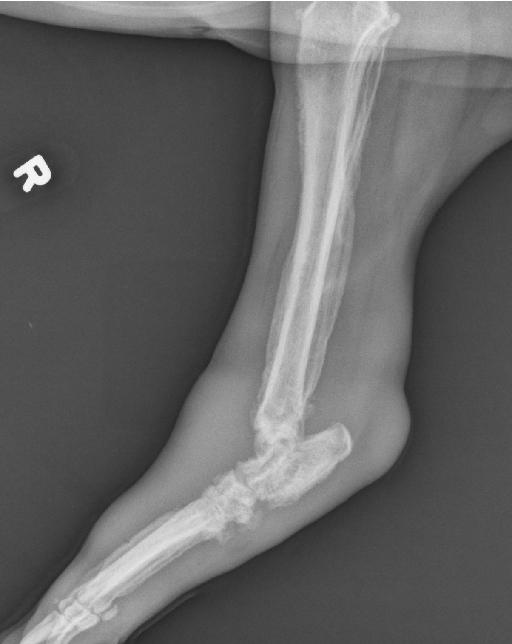
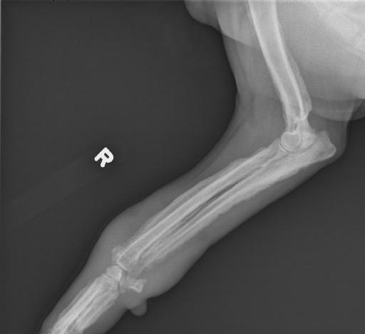
There is smooth palissading perisoteal proliferation noted on the distal aspect of the humeri, radii, and ulnae.
There are two ballistic fragments superimposed with the thorax. The liver is mildly enlarged, with rounded borders, and mild deviation of the gastric axis caudally.


Diagnosis
Pulmonary mass with dystrophic mineralization. Primary consideration is given to a neoplastic process. Trachebronchial lymphadenopathy. Pulmonary metastatic disease.
Hypertrophy osteopathy.
Mild unspecific hepatopathy. Ballistic fragments.
Pathology Report
FNAs of the mass were performed and a diagnosis of extraskelettal osteosarcoma.
Notes
Files