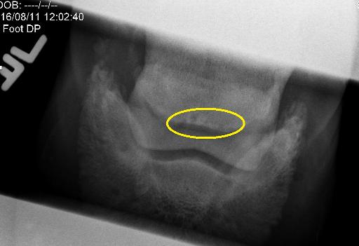
Equine navicular degeneration
Publication Date: 2016-08-15
Details
Service Radiology
Modality: Radiographs
Species: Equine
Area: Limb
History
16 year old American Quarter Horse. Suspected lame left forelimb, lameness eliminated after a palmar digital nerve block.
4 images
Findings
Orthogonal and oblique views were obtained. The toe is long and there is mild to moderate latero-medial imbalance. There is mild sclerosis of the navicular bone. Five radiolucent synovial invaginations are visible, most of which are wider and taller than normal with abnormal shapes. The largest synovial invagination is surrounded by sclerosis and is visible on the navicular skyline view.

Notes
Files