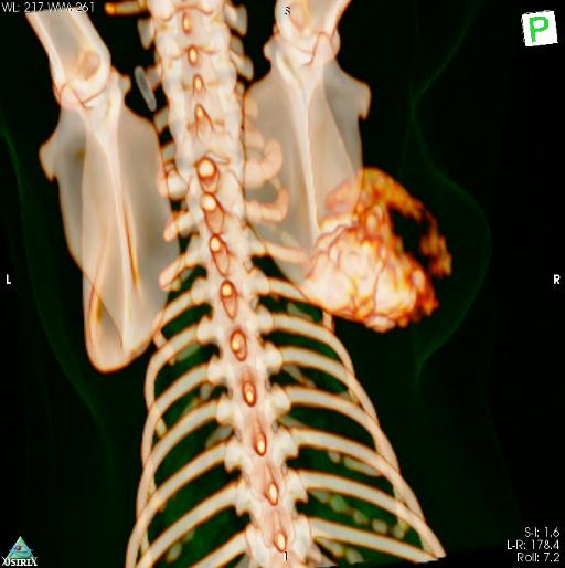
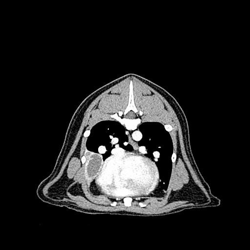
Scapula chondroblastic osteosarcoma
Publication Date: 2016-06-30
History
13 year old cat mixed breed cat
3 images
Findings
Orthogonal radiographs of the thorax are available for interpretation.
Centered at the level of the spine and proximo-lateral aspect of the right scapula, there is a large heterogeneously mineralized mass. This is associated with a moderate to large amount of surrounding soft tissue swelling. The margins of the scapular spine are no longer seen.
Overall the cardiovascular structures are considered to be within normal limits. An alveolar pattern is seen within the right middle lung lobe, this is most visualized on the VD projection. This results in a cranial mediastinal shift with the cardiac silhouette deviated towards the right side and partial border effacement of the cardiac silhouette. The remainder of the pulmonary parenchyma is considered to be within normal limits for the age of the patient.
Diagnosis
Agressive scapular mass, primary consideration is given to a neoplastic process such as chondrosarcoma, osteosarcoma or fibrosarcoma.
Pulmonary changes could represent chronic right middle lung lobe consolidation. However, underlying pulmonary disease cannot be ruled out.
No evidence of pulmonary metastasis radiographically.
Pathology Report
The scapular mass was diagnosed as a chondroblastic osteosarcoma. The mass was surgically removed.
On CT a cavitated mass was seen within the right middle lung lobe. This was diagnosed as a pulmonary abscess on FNAs, likely unrelated to the scapular mass.


Notes
Files