12.26.17
Publication Date: 2016-06-30
History
8 year old female spayed Shetland Sheepdog. Coughing.
3 images
Findings
Orthogonal radiographs of the thorax are available for interpretation.
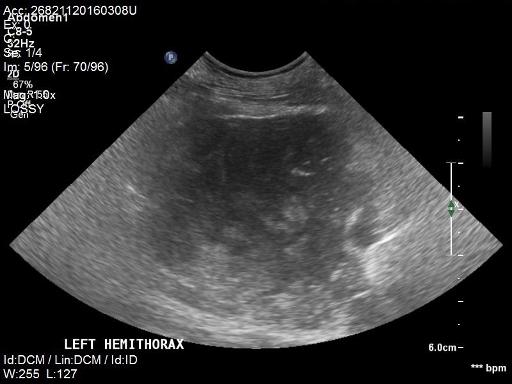
Spanning the first three ribs, within the cranial mediastin there is a well-defined irregularly marginated soft tissue mass. The mass displaces the trachea dorsally, and the pulmonary parenchyma caudally. Overall the mass is slightly more so on the left side and there is border effacement of the cranial left aspect of the cardiac silhouette.
The pulmonary parenchyma and cardiovascular structures are considered to be within normal limits. There is no radiographic evidence of pulmonary metastasis. The musculoskeletal system is overall within normal limits.
Incidentally there is a mild amount of soft tissue in the dorsal aspect of the caudal cervical trachea most consistent with a redudant tracheal membrane. There is also mild hepatomegaly with extension of the hepatic margins beyond the costal arch and caudal deviation of the gastric axis.
Diagnosis
Cranial mediastinal mass, primary consideration is given to a neoplastic process such as histiocytic sarcoma, thymoma,lymphoma less likely ectopic thyroid carcinoma. No evidence of pulmonary metastasis radiographically.
Redundant tracheal membranes.
Non-specific mild hepatomegaly. Consider benign hepatopathy vs less likely neoplastic process.
Pathology Report
The cranial mediastinal mass was diagnosed on FNAs with ultrasound as hystiocytic sarcoma.

Notes
Files