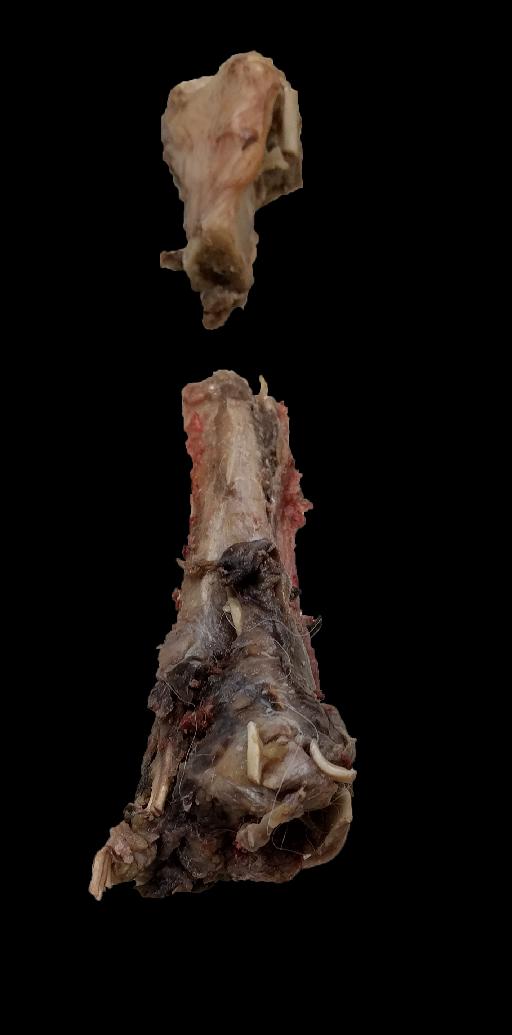
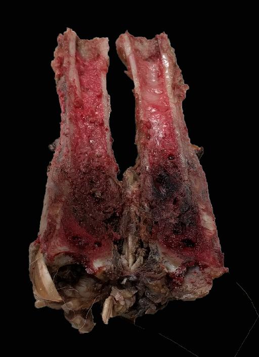
Pathologic fracture
Publication Date: 2016-05-03
Details
Service Anatomy
Modality: Radiographs
Species: Canine
Area: Head
4 images
Findings
Orthogonal and oblique radiographs centered on the left tarsus are available for interpretation.
At the level of the distal tibial diaphysis there is a moderate amount of soft tissue swelling. Affecting both the tibia and the fibula there is bicortical discontinuity. The fracture lines are oriented proximo-medial to disto-lateral. The distal tibial fragment is mildly displaced caudally and there is mild overiding of the ossseous fragments. There is minimal displacement at the level of the fibula.
Overall the distal tibia is more radiolucent than expected with thinning of the cortices. There is also a mild to moderate amount of new periosteal proliferation on the caudal aspect of the distal tibial diaphysis associated with a long zone of transition and a Codman's triangle.
It is difficult to evaluate the osseous quality of the fibula due to the superimposition but there is no definitive evidence of lysis radiographically
Diagnosis
Pathologic fracture of the left distal tibia and associated soft tissue swelling.
Secondary vs. less likely pathologic fracture of the left distal fibula.
Notes
Files

