Pulmonary carcinoma accessory lunge lobe
Publication Date: 2016-04-10
History
9 year old Bichon Frise, male castrated. Increased respiratory effort since last night, non productive cough, pain on abdominal palpation
6 images
Quizz
- Where is the mass arising from ?Diaphragm
No :)Accessory lung lobe
Yes :)Left caudal lung lobe
No :)Right caudal lung lobe
No :)Liver
No :)
Findings
Orthogonal radiographs of the thorax and abdomen are available for interpretation.
Thorax: In the caudal aspect of the thorax on midline there is a large well defined soft tissue mass. The mass displaces laterally both caudal lung lobes on the VD projection. On the VD projection there is splitting of the primary bronchi which are deviated dorsally on the lateral projection. Ventrally the margins of the diaphragm are indistinct, there is also partial border effacement with the cardiac silhouette. The caudal vena cava is completely effaced. There are very faint pleural fissure lines separating the lung lobes on the left side on the VD projection, a similar faint fissure line is noted between the right middle and right caudal lung lobe. The remainder of the pulmonary parenchyma is within normal limits. The cardiac silhouette is within normal limits for the age, breed and body condition score of the patient.
Abdomen: There is very mild loss of serosal detail in the cranioventral abdomen. There is a moderate amount of feces within the lumen of the colon. The margins of the liver extend beyond the costal arch and are mildly blunted. The kidneys are difficult to individualize but are small and contain a small amount of punctate mineralization. Between the right and left lateral projection of the abdomen there is significant variation in the size of the urinary bladder consistent with micturition. Incidentally there is mild degenerative joint disease affecting one of the stifle.
Diagnosis
Large pulmonary mass likely arising from the accessory lung lobe. Primary consideration is given to a neoplastic process such as bronchogenic carcinoma. No evidence of pulmonary metastasis. Scant amount of pleural effusion
Hepatomegaly and mild abdominal effusion. This is unspecific but is likely secondary to compression of the caudal vena cava by the pulmonary mass.
Bilateral chronic renal changes and presence of dystrophic mineralization
Incidental stifle arthritis.
Pathology Report
The mass was diagnosed as a pulmonary carcinoma with presence of mineralization and necrosis based on FNAs.


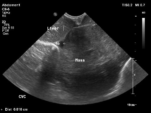
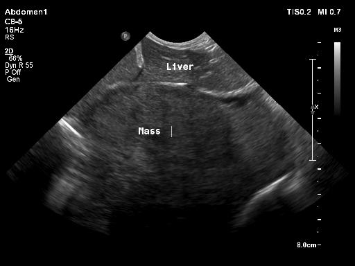
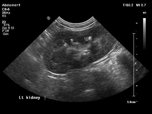
On the ultrasound the mass was visualized adjacent to the diaphragm. The caudal vena cava was compressed and there was enlargement of the hepatic veins. There was presence of free abdominal fluid and the chronic renal changes were confirmed with presence of nephroliths and loss of corticomedullary definition of the kidneys.
Acknowledgment
These images are courtesy of the University of Tennessee.