14y Chihuahua presenting for possible gall bladder mucocele
Publication Date: 2016-02-01
History
14y Chihuahua presenting for possible gall bladder mucocele
**Compare to the case of the week on 01/19/2016
3 images
Findings
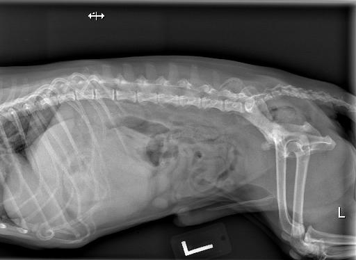
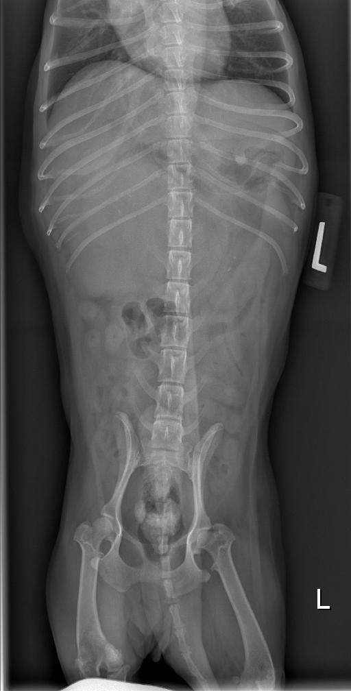
There is a decrease in peritoneal detail within the cranioventral abdomen due to a large soft tissue mass effect on the right side. This mass is homogeneously soft tissue, well defined, and round in shape. This mass appears contiguous with the caudal aspect of the liver on both lateral projections and the VD projection. On the right lateral projection, there is at least one intestinal loop ventral to this soft tissue mass. The descending duodenum is displaced medially on the VD. The intestinal tract is caudally displaced. There is no ventral displacement of the colon.
Two kidneys are visualized on the lateral and VD projections. There is multifocal, ill defined mineralization associated with the both kidneys that is best visualized on the VD projection. The urinary bladder is moderately distended with fluid. There are multiple large, stellate shaped mineralized structures within the bladder neck.
Diagnosis
Soft tissue mass within the right cranioventral abdomen. Given the patient's age, neoplasia is considered the most likely differential (hepatocellular carcinoma, hemangiosarcoma). Tissue of origin is most likely the right side of the liver. Spleen cannot be ruled out, but is considered less likely. Abscess or granuloma formation may also be considered, but are considered less likely.
A right sided liver mass was confirmed on ultrasound.
Multiple, mineralized urinary stones. Bilateral nephroliths.
Notes
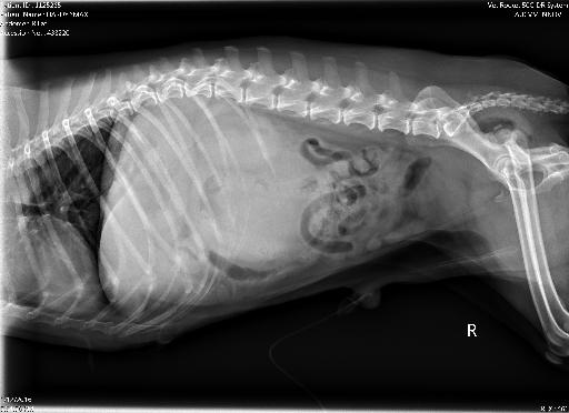
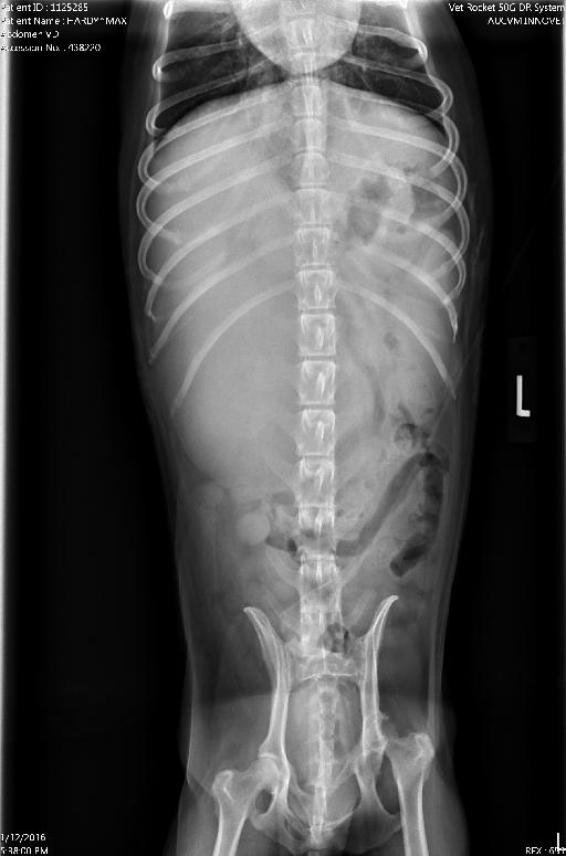
Did you compare this to the case from 01/19/2016, our 17y Jack Russell?
I thought these two cases were interesting to compare side by side. The VD images have a very similar appearing mass effect; despite the masses originating from different tissues.
Also, note the difference between the displacement of the bowel on the lateral projections. Today's case, the colon appears to be in its normal location due to the mass being ventrally located in the peritoneal space. The previous case, the colon is ventrally displaced, due to the mass originating in the retroperitoneal space.
Today's patients are the first two images.


Files