6y FS Great Dane The patient presented to the referring veterinarian for right pelvic limb lameness. The referring veterinarian had palpated a luxating right coxofemoral joint and had reduced it. However, the patient began having more difficulties rising, and ataxia in both hind limbs, and was referred to the neurology service.
Publication Date: 2015-10-25
History
6y FS Great Dane The patient presented to the referring veterinarian for right pelvic limb lameness. The referring veterinarian had palpated a luxating right coxofemoral joint and had reduced it. However, the patient began having more difficulties rising, and ataxia in both hind limbs, and was referred to the neurology service.
On presentation to neurology - the patient's neurologic status was localized to L3-S3.
Pelvis and spinal radiographs were recommended.
4 images
Findings
Spine: There is patchy lysis and moderate sclerosis noted in the spinous process of L5 extending through the lamina and the pedicles. The caudal articular facet may also be affected. The intervertebral foramen at L5-L6 has increased opacity and it is smaller than the adjacent intervertebral foramina.
Pelvis: Mild degenerative changes are associated with both coxofemoral joints.There is mild decreased coverage of the right coxofemoral joint.

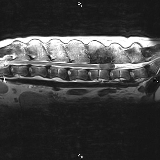
This is a sagittal T2-weighted MR image of the lumbar spine which shows marked destruction of L5, in addition to a large mass within the vertebral foramen.
Diagnosis
Lytic lesion associated with the vertebral body/spinous process of L5.
Differentials: Primary versus metastatic neoplasia of the bone. Bacterial or fungal osteomyelitis are considered less likely.
Files