Pulmonary mass+bullae
Publication Date: 2015-05-11
3 images
Findings
Orthogonal radiographs of the thorax are available for interpretation.
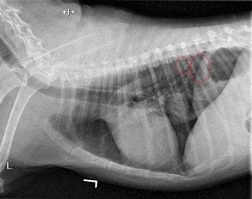
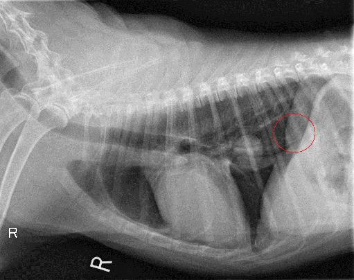
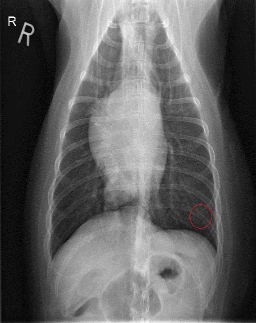
There is a well-defined single rounded soft tissue mass measuring about 40 mm in diameter visualized on the lateral projection just dorsal to the caudal vena cava caudal to the mainstem bronchi. There is mild ventral deviation of the caudal vena cava on the left lateral projection. Furthermore in the caudo dorsal lung field there are 2 well-defined thin walled gas filled structures visualized. The cranial one measures about 14 mm in diameter the caudal one measures about 25 mm in diameter. Similar structure is visualized superimposed with the diaphragmatic crus just dorsal to the caudal vena cava and measures about 21 mm on the right lateral projection. The cardiovascular structures are considered to be within normal limits.
Impression: 1. Single soft tissue mass superimposed with the accessory lung lobe. Primary consideration is given to a neoplastic process such as primary neoplasia or pulmonary metastasis. 2. The described above gas filled thin-walled structures are most consistent with pulmonary bullae.
Notes
Files


