Megaesophagus
Publication Date: 2015-04-20
History
1 year old dachshund with history of regurgitation.
2 images
Findings
Orthogonal radiographs of the thorax are available for interpretation.
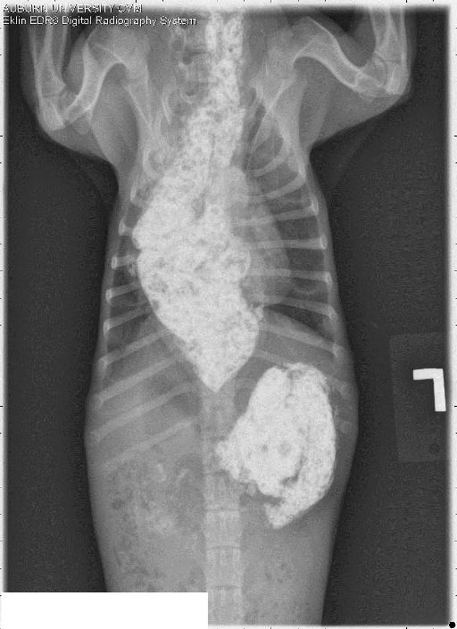
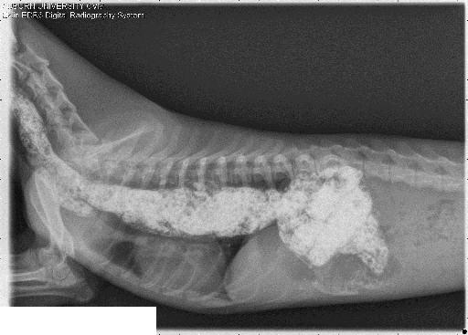
Within the dorsal thorax from the level of the caudal cervical area and to the level of the caudal thoracic area, the esophagus is gas distended. At its maximum, the esophagus measures approximately 25 mm in height. This results in ventral deviation and compression of the trachea and cardiac silhouette. On the ventral dorsal projection, the esophagus is mostly visualized superimposed with the right hemithorax. The pulmonary parenchyma and cardiovascular structures are considered to be within normal limits There is a small amount of radiopaque material noted within the lumen of the stomach. Impression: 1. Changes consistent with megaesophagus. 2. No evidence of aspiration pneumonia. For further evaluation and if clinically indicated barium oesophagram could be performed
Diagnosis
Megaesophagus
Additional imaging
A mixture of barium and food was fed the patient. The previously largely distended esophagus is visualized. There is no radiographic evidence of peristaltic waves. The changes described above remain consistent with the diagnosis of megaesophagus. Esophageal dysmotility is very likely.


Notes
Megaesophagus is the most common cause of regurgitation in dogs and the most frequently reported motility disorder affecting the canine esophagus. It is associated with reduced muscle tone and peristaltic activity and can lead to transport disorders. Megaesophagus can be segmental or generalized and occurs secondary to diseases of the neuromuscular junction[myasthenia gravis], striated muscle [myositis], peripheral nerves [polyneuropathy] or central nervous system [inflammatory, toxic and neoplastic].
Thrall, 6th edition page 510
Files