Esophageal foreign body
Publication Date: 2015-03-01
History
11 months old GSD intact male dog, vomiting/regurgitation
3 images
Findings
Orthogonal radiographs of the thorax are available for interpretation.
Within the lumen of the esophagus, at the level of the 3rd, 4th and 5th rib there is a radiopaque granular foreign body. Within the foreign body numerous rectangularly shaped osseous structures are appreciated. The cervical esophagus and the cranial thoracic esophagus adjacent to the foreign body is gas filled. At the level of the 4th rib, there is ventral deviation of the trachea. The caudal esophagus from the level of the 6th rib to the level of the diaphragmatic crus is partially fluid filled. Radiographically there is no evidence of pneumomediastinum on current examination.
An alveolar pattern affecting the right middle lung lobe as characterized by the presence of numerous air bronchograms, partial silhouetting of the cardiac silhouette and lobar sign is noted.
The caudal vena cava and pulmonary vasculature are on the lower end of normal limits.
Diagnosis
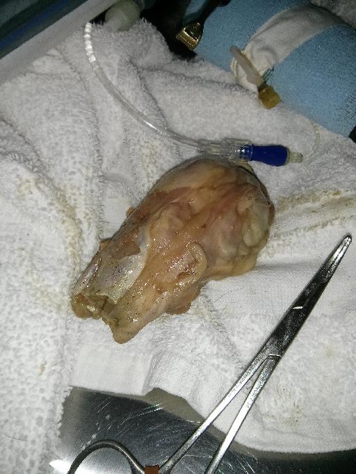
Impression: 1. Foreign body within the lumen of the thoracic esophagus at the level of the heart base. Based on the history of the patient is most consistent with a turkey's neck. 2. Alveolar pattern affecting the right middle lung lobe, most consistent with aspiration pneumonia. 3. Hypovolemia.
Gross

Notes
Files
References
- Esophageal FB are more common in dogs than cats. Foreign bodies are most often located at the thoracic inlet, base of the heart, or just cranial to the diaphragm, sites where the esophagus is limited in its ability to distend. A nonopaque esophageal FB may appear similar to an esophageal neoplasm, an esophageal abscess, mediastinal mass, paraesophageal hernia, or lung mass. Contraindications for a barium swallow include survey radiographic evidence of pneumothorax, pneumomediastinum, and pleural fluid which are signs of potential esophageal perforation. Thrall 6th edition, pages 511-513