False mets
Publication Date: 2015-01-21
Details
Service Radiology
Modality: Radiographs
Species: Canine
Area: Head
3 images
Findings
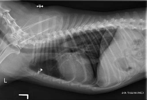
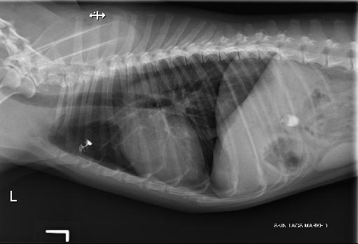
On the left lateral projection at the level of the 3rd intercostal space, in the ventral thorax a well defined soft tissue nodule measuring about 4mm is noted. This nodule is however not visualized on any other projection. On the ventrodorsal projection a rounded structure is superimposed with the spine of the left scapula and measures about 7mm.
Further imaging
The student on the case indicated the presence of a skin mass in the area. The skin lump was coated with barium, and radiographs repeated. The skin lump was the cause of the shadow noted superimposed with the lung parenchyma.


- Radiographically, what is the smallest size of a detectable pulmonary metastases ?1-3mm
Wrong :D3-5mm
Wrong :D5-7mm
Wrong :D7-9mm
Right !! Thrall 6th edition, pages 621
Diagnosis
Final diagnosis: Skin lump mimicking a potential pulmonary metastasis.
Notes
Files